Bone Grafting
Bone Grafting Overview:
- Socket Preservation (Minor Bone Grafting)
- Major Bone Grafting
- The Importance of Teeth for Jaw Bone Health
- Potential Consequences of Tooth and Jawbone Loss
- Reasons for Jawbone Loss and Deterioration
- About Bone Grafting
- Types of Bone Grafts
- Bone Graft Substitutes
- Socket Preservation
- Sinus Lift Procedure
- Am I a Candidate for a Sinus Lift Procedure?
- How is a Sinus Lift Accomplished?
- Ridge Augmentation/Expansion
What is bone grafting?
>> Request A Consultation
>> After Care Instructions for Bone Grafting
Over a period of time, the jawbone associated with missing teeth atrophies, or is reabsorbed. This often leaves a condition in which there is poor quality and quantity of bone suitable for placement of dental implants. In these situations, most patients are not candidates for placement of dental implants.
Today, we have the ability to grow bone where needed. This not only gives us the opportunity to place implant of proper length and width, it also gives us a chance to restore functionality and esthetic appearance.
Are you a candidate for bone grafting?
Dr. Hammond or Dr. Haralson can use bone grafting to help place implants for patients with inadequate bone structure. Contact us today to learn more!
Socket Preservation (Minor Bone Grafting)
The most common bone grafting procedure is done to preserve the height and width of the bone when the tooth is extracted in preparation for an implant placement. It is very important when removing the tooth in an area of future implant placement to remove it with great care and to maintain most of the bone surrounding the tooth. This is why you want the procedure to be performed by a surgeon experienced not just in removing teeth, but in removing teeth with preservation of the surrounding bone. In addition to extraction with bone preservation, it is important to graft the socket with bone to provide a scaffold for bone growth within the socket which preserves width and height. Drs. Hammond or Haralson performs thousands of extractions with bone preservation in addition to socket preservation every year. He is an expert at this procedure and will talk to you about this during your consultation.Major Bone Grafting
Bone grafting can repair implant sites with inadequate bone structure due to previous extractions, gum disease, or injuries. Although grafting procedures are not needed now as much as they were in the past because of CT-guided surgery and zygomatic implants, there are still several indications. The bone is either obtained from a tissue bank or your own bone is taken from the jaw, hip, or tibia (below the knee.) Sinus bone grafts are also performed to replace bone in the posterior upper jaw. In addition, special membranes may be utilized that dissolve under the gum and protect the bone graft and encourage bone regeneration. This is called guided bone regeneration or guided tissue regeneration. Drs. Hammond or Haralson has done hundreds of grafting cases and is an expert in this field.The Importance of Teeth for Jaw Bone Health
When one or more teeth are missing, it can lead to jawbone loss at the site of the gap. This loss of jawbone can develop into additional problems, both with your appearance and your overall health. You may experience pain, problems with your remaining teeth, and altered facial appearance, and eventually even the inability to speak and eat normally.
In that same way that muscles are maintained through exercise, bone tissue is maintained by use. Natural teeth are embedded in the jawbone, and stimulate the jawbone through activities such as chewing and biting. When teeth are missing, the alveolar bone, or the portion of the jawbone that anchors the teeth in the mouth, no longer receives the necessary stimulation, and begins to break down, or resorb. The body no longer uses or “needs” the jawbone, so it deteriorates and goes away.
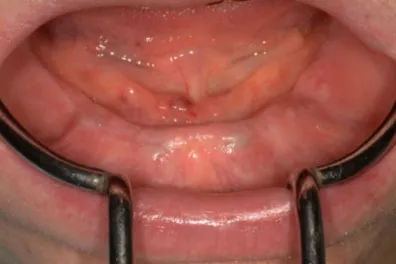
Severe resorption of the lower ridge following teeth extractions. The patient complains of inability to keep her denture in place.
Potential Consequences of Tooth and Jawbone Loss
- Problems with remaining teeth, including, misalignment, drifting, loosening and loss
- Collapsed facial profile
- Limited lip support
- Skin wrinkling around the mouth
- Distortion of other facial features
- Jaw pain, facial pain, and headaches
- Difficulty speaking and communicating
- Inadequate nutrition as a result of the inability to chew properly and painlessly
- Sinus expansion
Reasons for Jawbone Loss and Deterioration
The following are the most common causes for jawbone deterioration and loss that may require a bone grafting procedure:
Tooth Extractions:
When an adult tooth is removed and not replaced, jawbone deterioration may occur. Natural teeth are embedded in the jawbone, and stimulate the jawbone through activities such as chewing and biting. When teeth are missing, the alveolar bone, or the portion of the jawbone that anchors the teeth in the mouth, no longer receives the necessary stimulation, and begins to break down, or resorb. The body no longer uses or “needs” the jawbone, so it deteriorates and goes away.
The rate the bone deteriorates, as well as the amount of bone loss that occurs, varies greatly among individuals. However, most lost occurs within the first eighteen months following the extraction, and continues throughout life.
Periodontal Disease:
Periodontal diseases are ongoing infections of the gums that gradually destroy the support of your natural teeth. Periodontal disease affects one or more of the periodontal tissues: alveolar bone, periodontal ligament, cementum, or gingiva. While there are many diseases which affect the tooth-supporting structures, plaque-induced inflammatory lesions make up the majority of periodontal issues, and are divided into two categories: gingivitis and periodontitis. While gingivitis, the less serious of the diseases, may never progress into periodontitis, it always precedes periodontitis.
Dental plaque is the primary cause of gingivitis in genetically-susceptible individuals. Plaque is a sticky colorless film, composed primarily of food particles and various types of bacteria, which adhere to your teeth at and below the gum line. Plaque constantly forms on your teeth, even minutes after cleaning. Bacteria found in plaque produce toxins or poisons that irritate the gums. Gums may become inflamed, red, swollen, and bleed easily. If this irritation is prolonged, the gums separate from the teeth causing pockets (spaces) to form. If daily brushing and flossing is neglected, plaque can also harden into a rough, porous substance known as calculus (or tartar). This can occur both above and below the gum line.
Periodontitis is affected by bacteria that adhere to the tooth’s surface, along with an overly aggressive immune response to these bacteria. If gingivitis progresses into periodontitis, the supporting gum tissue and bone that holds teeth in place deteriorates. The progressive loss of this bone, the alveolar, can lead to loosening and subsequent loss of teeth.
Are you a candidate for bone grafting?
Drs. Hammond or Haralson can use bone grafting to help place implants for patients with inadequate bone structure. Contact us today to learn more!
Dentures/Bridgework:
Unanchored dentures are placed on top of the gum line, and therefore do not provide any direct stimulation to the underlying alveolar bone. Over time, the lack of stimulation causes the bone to resorb and deteriorate. Because this type of denture relies on the bone to hold them in place, people often experience loosening of their dentures and problems eating and speaking. Eventually, bone loss may become so severe that dentures cannot be held in place even with strong adhesives, and a new set may be required. Proper denture care, repair, and refitting are essential to maintaining oral health.
Some dentures are supported by anchors, which do help adequately stimulate, and therefore preserve bone.
With bridgework, the teeth on either side of the appliance provide sufficient stimulation to the bone, but the portion of the bridge that spans the gap where the teeth are missing receives no direct stimulation. Bone loss can occur in this area.
By completing a bone graft procedure, Drs. Hammond or Haralson is now able to restore bone function and growth, thereby halting the effects of poor denture care.
Trauma:
When a tooth is knocked out or broken to the extent that no biting surface is left below the gum line, bone stimulation stops, which results in jaw bone loss. Some common forms of tooth and jaw trauma include: teeth knocked out from injury or accident, jaw fractures, or teeth with a history of trauma that may die and lead to bone loss years after the initial trauma.
A bone grafting procedure would be necessary to reverse the effects of bone deterioration, restoring function and promoting new bone growth in traumatized areas.
Misalignment:
Misalignment issues can create a situation in the mouth where some teeth no longer have an opposing tooth structure. The unopposed tooth can over-erupt, causing deterioration of the underlying bone.
Issues such as TMJ problems, normal wear-and-tear, and lack of treatment can also create abnormal physical forces that interfere with the teeth’s ability to grind and chew properly. Over time, bone deterioration can occur where bone is losing stimulation.
Osteomyelitis:
Osteomyelitis is a type of bacterial infection in the bone and bone marrow of the jaw. The infection leads to inflammation, which can cause a reduction of blood supply to the bone. Treatment for osteomyelitis generally requires antibiotics and removal of the affected bone. A bone graft procedure may then be required to restore bone function and growth lost during removal.
Tumors:
Benign facial tumors, though generally non-threateningly, may grow large and require removal of a portion of the jaw. Malignant mouth tumors almost always spread into the jaw, requiring removal of a section of the jaw. In both cases, reconstructive bone grafting is usually required to help restore function to the jaw. Grafting in patients with malignant tumors may be more challenging because treatment of the cancerous tumor generally requires removal of surrounding soft tissue as well.
Developmental Deformities:
Some conditions or syndromes known as birth defects are characterized by missing portions of the teeth, facial bones, jaw or skull. Drs. Hammond or Haralson may be able to perform a bone graft procedure to restore bone function and growth where it may be absent.
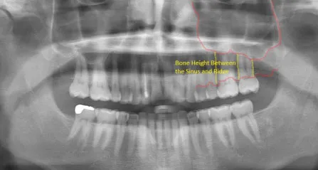
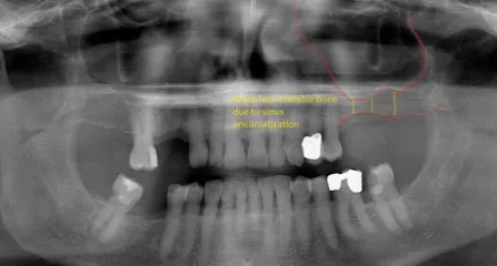
Sinus Deficiencies:
When molars are removed from the upper jaw, air pressure from the air cavity in the maxilla (maxillary sinus), causes resorption of the bone that formerly held the teeth in place. As a result, the sinuses become enlarged, a condition called pneumatization of the sinus.
Normal Maxillary Sinus
Pneumatized Maxillary Sinus
About Bone Grafting
What is Bone Grafting?
Over a period of time, the jawbone associated with missing teeth atrophies and is reabsorbed. This often leaves a condition in which there is poor quality and quantity of bone suitable for placement of dental implants. In these situations, most patients are not candidates for placement of dental implants.
With bone grafting, we now have the opportunity to not only replace bone where it is missing, but also the ability to promote new bone growth in that location! This not only gives us the opportunity to place implants of proper length and width, it also gives us a chance to restore functionality and esthetic appearance.
Types of Bone Grafts
Autogenous Bone Grafts:
Autogenous bone grafts, also known as autografts, are made from your own bone, taken from somewhere else in the body. The bone is typically harvested from the chin, jaw, lower leg bone, hip, or the skull. Autogenous bone grafts are advantageous in that the graft material is live bone, meaning it contains living cellular elements that enhance bone growth.
However, one downside to the autograft is that it requires a second procedure to harvest bone from elsewhere in the body. Depending on your condition, a second procedure may not be in your best interest.
Allogenic Bone:
Allogenic bone, or allograft, is dead bone harvested from a cadaver, then processed using a freeze-dry method to extract the water via a vacuum. Unlike autogenous bone, allogenic bone cannot produce new bone on its own. Rather, it serves as a framework or scaffold over which bone from the surrounding bony walls can grow to fill the defect or void.
Xenogenic Bone:
Xenogenic bone is derived from non-living bone of another species, usually a cow. The bone is processed at very high temperatures to avoid the potential for immune rejection and contamination. Like allogenic grafts, xenogenic grafts serve as a framework for bone from the surrounding area to grow and fill the void.
Both allogenic and xenogenic bone grafting are advantageous in that they do not require a second procedure to harvest your own bone, as with autografts. However, because these options lack autograft’s bone-forming properties, bone regeneration may take longer than with autografts, with a less predictable outcome.
Bone Graft Substitutes
As a substitute to using real bone, many synthetic materials are available as a safe and proven alternative, including:
Demineralized Bone Matrix (DBM)/Demineralized Freeze-Dried Bone Allograft (DFDBA):
This product is processed allograft bone, containing collagen, proteins, and growth factors that are extracted from the allograft bone. It is available in the form of powder, putty, chips, or as a gel that can be injected through a syringe.
Graft Composites:
Graft composites consist of other bone graft materials and growth factors to achieve the benefits of a variety of substances. Some combinations may include: collagen/ceramic composite, which closely resembles the composition of natural bone, DBM combined with bone marrow cells, which aid in the growth of new bone, or a collagen/ceramic/autograft composite.
Bone Morphogenetic Proteins:
Bone morphogenetic proteins (BMPs) are proteins naturally produced in the body that promote and regulate bone formation and healing.
Synthetic materials also have the advantage of not requiring a second procedure to harvest bone, reducing risk and pain. Each bone grafting option has its own risks and benefits. Drs. Hammond or Haralson will determine which type of bone graft material is right for you.
Socket Preservation
What is a Socket Preservation?
Socket Preservation is a common surgical procedure often performed following a tooth extraction to help recreate the natural contour of the gums and jaw that may have been lost due to bone loss as a result of a tooth extraction, or for another reason.
The alveolar ridge of the jaw is the bone that surrounds the roots of teeth. When a tooth is removed, an empty socket is left in the alveolar ridge bone. Usually this empty socket will heal on its own, filling with bone and tissue. Sometimes when a tooth is removed, the bone surrounding the socket breaks, and it is unable to heal on its own. The previous height and width of the socket will continue to deteriorate.
Rebuilding the original height and width of the alveolar ridge is not medically necessary, but may be required for dental implant placement, or for aesthetic purposes. Dental implants require bone to support their structure, and socket preservation can help rebuild this bone to accommodate the implant.
How is the Socket Preservation Surgery Accomplished?
Socket preservation is accomplished by placing bone graft material in the tooth socket. It is often done immediately after the tooth is removed, to avoid the need for a second procedure later. Next, the gum tissue is placed over the socket and secured with sutures. Drs. Hammond or Haralson may choose to use a space-maintaining product over the top of the graft to help restore the height and width of the space created by the tooth and bone loss, and into which new bone should grow. Once the socket has healed, the alveolar ridge can be prepared for dental implant placement.
Socket preservation is typically performed in Drs. Hammond or Haralson‘s office under local anesthesia. Some patients may also request sedative medication in addition.
Sinus Lift Procedure
The maxillary sinuses are behind your cheeks and on top of the upper teeth. These sinuses are empty, air-filled spaces. Some of the roots of the natural upper teeth extend up into the maxillary sinuses. When these upper teeth are removed, there is often just a thin wall of bone separating the maxillary sinus and the mouth. Dental implants need bone to hold them in place. When the sinus wall is very thin, it is impossible to place dental implants in this bone.
Normal Maxillary Sinus
Pneumatized Maxillary Sinus
There is a solution and it’s called a sinus graft or sinus lift graft. The surgery is performed by entering the sinus from where the upper teeth used to be. The sinus membrane is then lifted upward and donor bone is inserted into the floor of the sinus. Keep in mind that the floor of the sinus is the roof of the upper jaw. After several months of healing, the bone becomes part of the patient’s jaw and dental implants can be inserted and stabilized in this new sinus bone.
The sinus graft makes it possible for many patients to have dental implants when years ago there was no other option other than wearing loose dentures.
If enough bone between the upper jaw and the bottom of the sinus is available to stabilize the implant well, sinus augmentation and implant placement can sometimes be performed as a single procedure. If not enough bone is available, the sinus augmentation will have to be performed first, then the graft will have to mature for several months, depending upon the type of graft material used. Once the graft has matured, the implants can then be placed.
The key to a successful and long-lasting dental implant is the quality and quantity of jawbone to which the implant will be attached. If bone loss has occurred due to injury or periodontal disease, a sinus augmentation can raise the sinus floor and allow for new bone formation. A sinus lift is one of the most common bone grafting procedures for patients with bone loss in the upper jaw. The procedure seeks to grow bone in the floor of the maxillary sinus above the bony ridge of the gum line that anchors the teeth in the upper jaw. By strengthening and growing bone in this location, dental implants can be placed and secured in the new bone growth.
Am I a Candidate for a Sinus Lift Procedure?
A sinus lift may be necessary if you:
- are missing more than one tooth in the back of your jaw.
- are missing a significant amount of bone in the back of your jaw.
- are missing teeth due to a birth defect or condition.
- are missing most of the maxillary teeth, but require support for dental implants.
How is a Sinus Lift Accomplished?
In the most common sinus augmentation procedure, a small incision is made on the premolar or molar region to expose the jawbone. A small opening is cut into the bone, and the membrane lining the sinus is pushed upward. The underlying space is filled with bone grafting material, either from your own body or from a cadaver. Sometimes, synthetic materials that can imitate bone formation are used. After the bone is implanted, the incision is stitched up and the healing process begins. After several months of healing, the bone becomes part of the patient’s jaw and dental implants can be inserted and stabilized in this new sinus bone.
If enough bone between the upper jaw ridge and the bottom of the sinus is available to stabilize the implant well, sinus augmentations and implant placement can sometimes be performed as a single procedure. If not enough bone is available, the sinus augmentation will have to be performed first, then the graft will have to mature for several months, depending upon the type of graft material used. Once the graft has matured, the implants can be placed.
The sinus graft makes it possible for many patients to have dental implants when years ago there was no other option besides wearing loose dentures.
A sinus augmentation is generally performed at Drs. Hammond or Haralson‘s office , under intravenous sedative medication as well.
Ridge Augmentation/Expansion
In severe cases, the ridge has been reabsorbed and a bone graft is placed to increase ridge height and/or width. This is a technique used to restore the lost bone dimension when the jaw ridge gets too thin to place conventional implants. In this procedure, the bony ridge of the jaw is literally expanded by mechanical means. Bone graft material can be placed and matured for a few months before placing the implant.